

Medical Imaging Technologies
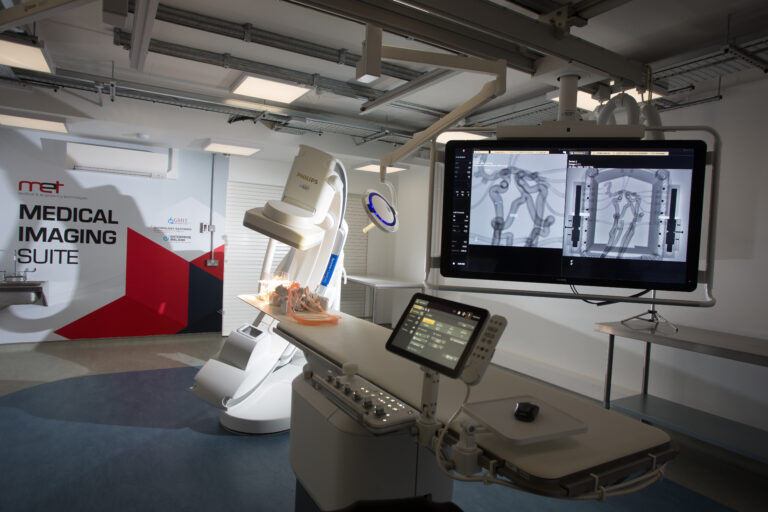
The Philips Azurion 7 M20 digital fluoroscope in the purpose-built Medical Imaging Suite consititues an outstanding resource for the MedTech industry and academic and clinical researchers. This fluoroscope is optimised for vascular, non-vascular, cardiovascular and neuro applications and with its high resolution and large field of view it offers superb image quality to evaluate small details and vessels with clarity.
While complimenting the MET Gateway’s existing expertise in medical imaging, anatomical modelling and physiological replication, this state-of-the-art medical testing facility for companies developing next generation medical technologies will enable our industry, research and academic partners access to equipment not readily available outside of a clinical environment to design, test and demonstrate features of their new technologies.
Overall, the Medical Imaging Suite positions the MET Gateway’s fluoroscopy capabilities to evolve from 2D traditional fluoroscopy offering to 3D augmented state-of-the-art hospital fluoroscopy and data post-processing. Additionally, it’s standardised hardware and software platform provides access to a whole new generation of connected healthcare applications and technologies.
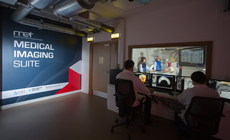
The Medical Imaging Suite can also be used for training of clinicians, staff and students in new imaging technologies, the interpretation of medical images and practice techniques as well as the evaluation and demonstration of new and emerging medical technologies.
The Philips Azurion 7 M20 digital fluoroscope key capabilities include…
- 3D Rotational Angiography allows the continuous 360 degrees visualisation of the region of interest and is performed during one rotational scan. Contrast agent is necessary to be injected in the vasculature or the cavities in the models.
- 3D Interventional Module (including the generation of a 3D geometry of the scanned object/model for R&D purposes) provides 3D Fluoroscopy data post processing i.e. data evaluation in terms of planning a procedure and taking measurements on the scanned models/objects/devices.
- XperCT Dual facilitates the acquisition of two scans at a defined interval, providing CT-like images, aiding in the assessment of soft tissue, bone structures and stent deployment. This is completed during one rotational scan and the data can be evaluated in the axial, sagittal and coronal planes like a normal CT dataset.
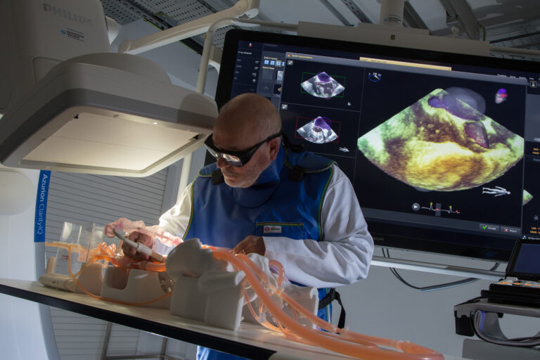
- 2D Quantitative Vascular Analysis feature which facilitates the quantitative assessment of aortic and peripheral vasculature, while VesselNavigator offers a complete live image guidance solution for endovascular procedures, providing an intuitive and continuous 3D road map to guide you through the vasculature. Alongside the HeartNavigator modules, this helps to train operators and/or to simulate (for R&D engineers and clinicians) the delivery of medical devices via realistic challenging tortuous vasculature with 3D visualisation feedback.
- SmartPerfusion which helps assess the changes in perfusion due to an intervention, be that vessel patency after an endovascular intervention or evaluating tumour perfusion after embolization. From a data analysis perspective, Smart Perfusion provides qualitative evaluation of improved/reduced perfusion in a model. This function would help to evaluate the success of an angioplasty/stenting procedure, for example, in animal tissue or artificial model.
- HeartNavigator is specifically designed to support a wide variety of structural heart disease procedures. It can automatically identify landmark points, provide automatic measurement and includes virtual device templates to allow for accurate sizing and virtual target positioning before device deployment.
- FlexVision XL HD 58” Monitor offers exceptional viewing, and can include displays from 3rd party equipment or other imaging modalities as desired, while Instant Parallel Working allows team members to view and analyse images in the control room, without interrupting an ongoing test in the examination room.
- A normal CT import function is also available on the system, with the capability to automatically position the C-arm at any scanning angle chosen on the CT dataset.
- MET’s Medical Imaging Suite also boasts Recording and Live-Streaming capabilities, facilitated by our MedInBox system. Customers can record activities within the suite while simultaneously live-streaming to off-site teams and clinical partners. The recording system is integrated with the Philips Azurion 7 M20 digital fluoroscope and the Philips EPIQ CVxi Ultrasound (via the EchoNavigator Software) and includes 3 cameras (2 stationary and 1 roving) and 5 head-sets. This combined facility allows users to record the fluoroscopic and ultrasound images in parallel with the activities on the bench and the audio within the room, ensuring every detail of your session is recorded in full.
This link provides further information on our equipment and capabilities.
If you would like further information or would like to book a tour, please contact us for advice and support.